SEED | 压力如何让造血干细胞提前变老 SEED | Stress pushes HSCs toward aging AI-assisted · reviewed
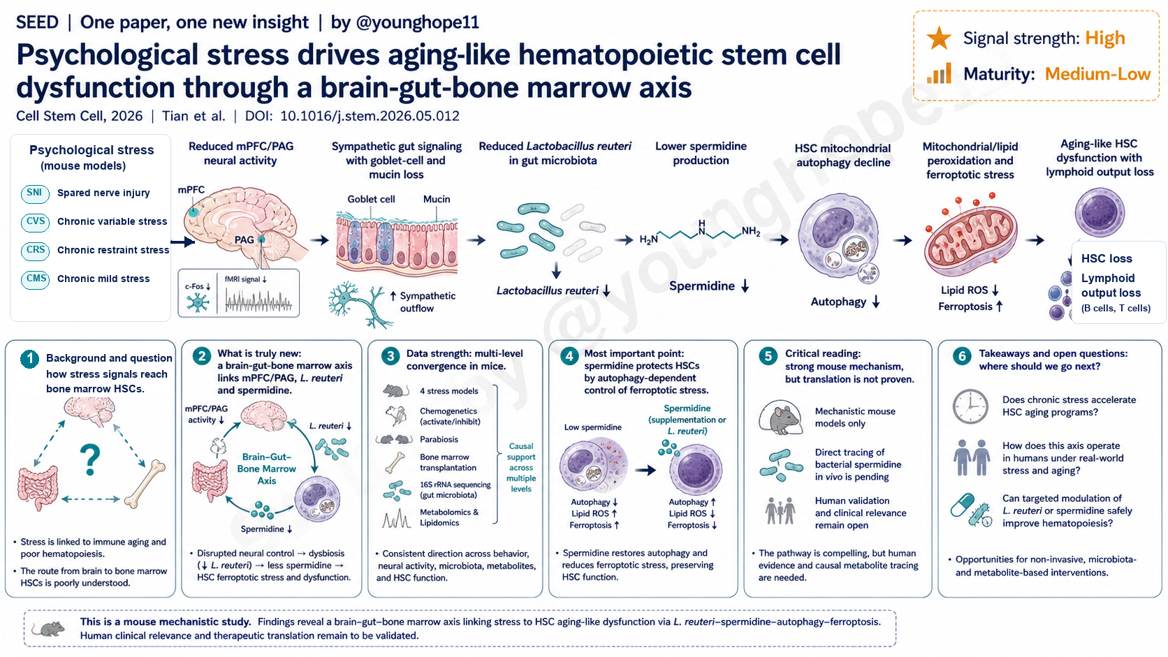
Sun Yat-sen University 的 Xiaobin Tian、Binghuo Wu、Keyue Yang、Ying Wang 与 Linjia Jiang、Meng Zhao 团队近期在 Cell Stem Cell 报道,在多种小鼠心理应激模型中,应激会通过 mPFC/PAG-交感神经-肠道菌群-骨髓轴降低 L. reuteri 与 spermidine,诱导 HSC 自噬下降、铁死亡压力上升和类衰老功能衰退,为理解压力如何进入造血系统提供了新的机制框架。
问题不是压力会不会影响免疫,而是它如何抵达 HSC
心理应激与心血管病、糖尿病、感染和肿瘤免疫反应下降有关,这一点并不新。更难的问题是:一个发生在中枢神经系统和情绪行为层面的状态,如何一路影响到骨髓深处的 hematopoietic stem cells。
这篇论文把问题放在 HSC 衰老上。HSC 衰老的典型特征包括自我更新能力下降、淋巴分化能力下降、髓系偏倚增加、线粒体压力和炎症程序增强。作者使用了四类小鼠应激模型:spared nerve injury、chronic variable stress、chronic restraint stress 和 chronic mild stress。前三者产生更强的行为和造血表型,CMS 表型较弱。
结果上,应激小鼠出现 HSC 数量下降、HSC cell death 上升、静息状态下降、myeloid-biased HSC 增加、common lymphoid progenitor 下降,以及外周 T/B 淋巴细胞输出下降。骨髓移植实验进一步显示,应激暴露后的 donor cells 在 primary recipients 中自我更新和淋巴重建能力受损,在 secondary recipients 中多谱系重建进一步下降。
因此,这篇文章不是泛泛地说“压力影响免疫”,而是把问题具体化为:心理应激是否会把年轻 HSC 推向一种功能上类似老化的状态,以及这条信号链是否能被拆成可干预的节点。
真正的新意:把脑区、肠菌和骨髓串成一条因果链
最有新意的部分,是作者没有停在炎症因子或肾上腺激素这个层面,而是把信号链向上追到脑区、向下追到肠道菌群和骨髓代谢物。
他们先用 c-Fos 和 resting-state fMRI 做区域活动定位,发现 SNI、CVS 和 CRS 等应激模型中 mPFC 和 PAG 活性下降。随后用 DREADD 化学遗传学分别抑制或激活脑区:抑制 mPFC 或 PAG 足以复现 HSC 丢失、my-HSC 增加和淋巴输出下降;在 SNI 小鼠中激活 mPFC 或 PAG 则能恢复 HSC 存活、数量和淋巴分化。
接着,作者用并体实验提示循环因子参与其中;用骨髓 extracellular fluid 代谢组发现 spermidine 在应激和 mPFC/PAG 抑制条件下下降;用 16S、菌株分离和细菌灌胃进一步把焦点收缩到一种高 spermidine 产生能力的 Lactobacillus reuteri strain。L.R.S. 补充可以恢复 serum 和 bone marrow spermidine,减少 HSC ferroptotic stress,并改善 HSC 自我更新和淋巴重建。
最后,神经路径的拆解也很关键。HSV tracing、vagotomy、celiac ganglionectomy、adrenalectomy、口服 6-OHDA 和肾上腺素受体阻断共同支持一个模型:mPFC/PAG 活性下降通过 RVLM-linked sympathetic pathway 改变小肠环境,造成 goblet cell 和 mucin 下降,进而降低 L. reuteri 和 spermidine,而不是主要通过 vagus nerve 或 HPA/SAM adrenal-derived signals。
数据强在多层扰动彼此对齐
这篇论文的数据强度来自多层证据的方向一致,而不是单个漂亮实验。行为学、脑区活动、化学遗传学、骨髓移植、并体、代谢组、16S、菌株分离、FMT、L.R.S. 灌胃、铁死亡抑制、GPX4 overexpression、Atg5 条件敲除和神经切断实验,都指向同一条轴。
在造血功能层面,作者不仅看了 HSC 数量,也看了 cell death、my-HSC、CLP、外周 T/B/myeloid output,以及 primary/secondary transplantation。这比单纯 phenotyping 更能支撑“自我更新和淋巴分化能力受损”的结论。
在机制层面,spermidine 的位置也经过了多次交叉验证。外源 spermidine 能救 mPFC/PAG 抑制和 SNI 模型中的 HSC 表型;低 spermidine-producing strain 不能复现 L.R.S. 的保护作用;polyamine transporter inhibition 会削弱 spermidine 的保护;spermine、putrescine、citalopram 或 fluoxetine 不能简单替代 spermidine。
在细胞死亡机制上,作者用 Ferrostatin-1、Liproxstatin-1、alpha-tocopherol、GPX4 overexpression、BODIPY-C11、lipidomics、TEM、MitoSOX、MitoPerOx、MitoTEMPO 等证据,把 HSC 损伤更具体地定位到 mitochondrial peroxidative stress 和 ferroptotic stress,而不是笼统的 apoptosis 或 necroptosis。
最重要的一点:spermidine 不是补品标签,而是自噬-铁死亡开关
这篇论文最重要的一点,是把 spermidine 从一个容易被大众化误读的“抗衰补充物”概念,拉回到 HSC 线粒体质量控制和铁死亡压力的具体机制中。
在作者的模型里,应激不是直接让 HSC “变老”,而是先关闭部分 stress-responsive brain circuit,改变肠道 sympathetic signaling,减少 L. reuteri 和 spermidine。spermidine 下降后,HSC 的 autophagy/mitophagy 活性下降,线粒体 peroxidative stress 和 lipid peroxidation 上升,最终让 HSC 对 ferroptosis 更敏感。
Atg5 条件敲除实验是这条机制链的关键。Atg5-deficient HSC 本身就有更高 mtROS、mitochondrial/lipid peroxidation 和 ferroptotic cell death;在缺乏 Atg5 的情况下,SNI 不能进一步加重很多表型,spermidine 也不能有效救回 HSC 功能。这说明 spermidine 的保护作用依赖一个仍然可用的 autophagy machinery。
因此,本文真正改变视角的地方在于:压力诱导的 HSC 类衰老,不只是“炎症多了”或“交感神经强了”,而是可以被读成一个从脑区活动到微生物代谢物,再到 HSC 线粒体自噬和铁死亡压力的质量控制崩塌。
怎样批判性地读:强机制不等于可立即转化
第一,所有核心证据都来自小鼠。SNI、CVS、CRS 和 CMS 是有用模型,但它们不等同于人类长期心理压力、抑郁、焦虑、慢性病压力或社会应激。尤其 SNI 带有神经损伤和疼痛成分,虽然作者用多模型和脑区操作增强了说服力,但人类外推仍需要谨慎。
第二,spermidine 的来源和去向还没有完全闭环。作者在 limitations 中明确提到,还需要 isotope-labeled tracing 来证明 bacterially associated spermidine 是否直接到达骨髓并作用于 HSC。现在的数据支持这条轴,但还不能把每一个分子流向都视为已经被直接追踪。
第三,spermidine rescue 并不完全。论文也承认,spermidine 补充不能完整恢复所有 aging-like HSC phenotypes,说明心理应激可能还通过其他 systemic metabolic remodeling、其他细胞类型或器官间接作用于 HSC。
第四,L. reuteri 和肠道菌群干预不能被直接翻译成“吃某种益生菌或 spermidine 就能抗压力衰老”。菌株、宿主背景、剂量、给药时机、安全性、长期生态影响和人类 HSC 终点都还没有解决。这篇文章给的是机制地图,不是临床建议。
下一步该追问:这条轴在人类压力和衰老中是否成立
最直接的下一步,是在人类慢性压力、抑郁、老化或免疫功能下降人群中,观察 mPFC/PAG 活性、交感肠道信号、L. reuteri abundance、serum/bone marrow spermidine、HSPC 状态和淋巴输出之间是否存在同向关系。即使不能立刻取得骨髓样本,也可以从外周免疫年龄、炎症、微生物组和代谢组开始。
第二步,是区分心理应激诱导的 HSC 类衰老和生理性 HSC 老化的重叠与差异。本文已经显示 stress-exposed HSC 与 aged HSC 在 stemness、lymphoid differentiation、myeloid program、oxidative stress 和 inflammatory programs 上相似,但 DNA damage response 和 adhesion signatures 变化较温和。这种“partial aging-like state”是否可逆,是很值得追踪的问题。
第三步,是做更精确的干预实验。比如,能否用特定 L. reuteri strain、spermidine analogue、autophagy activator、ferroptosis inhibitor、alpha-adrenergic modulation 或非侵入式脑区调控来保护 HSC?这些干预需要分清 prevention、reversal 和 resilience 三种不同目标。
最后,这条轴对 HSC Museum 或血液系统抗衰研究的启发很清楚:HSC 不只是骨髓内部的细胞,它会读取脑、肠、神经、菌群、代谢物和线粒体质量控制的整合状态。未来的 HSC aging 研究,可能越来越需要把神经免疫学、微生物组和干细胞代谢放在同一张图里看。
Yang 的信号评级:High
轴一,信号强度:High。 理由是这篇论文不是只给出相关性,而是在多个层面做了因果扰动:脑区抑制和激活、神经路径切断、FMT、L.R.S. 补充、spermidine 补充、Atg5 条件敲除、GPX4 overexpression 和铁死亡抑制,彼此支持同一条 brain-gut-bone marrow axis。
轴二,成熟度:Medium-Low。 理由是机制证据在小鼠中很完整,但临床成熟度还低。人类慢性压力、肠道菌群、spermidine 和 HSC 功能之间是否存在同样的因果链,还需要队列、干预和更直接的代谢物追踪验证。
一句话总结:这篇文章的核心价值,是把“压力让免疫变老”从一句宏观判断,拆成了 mPFC/PAG、交感肠道信号、L. reuteri、spermidine、自噬和铁死亡压力组成的一条可实验检验的机制链。
Xiaobin Tian, Binghuo Wu, Keyue Yang, Ying Wang, Linjia Jiang, Meng Zhao and colleagues at Sun Yat-sen University recently reported in Cell Stem Cell that, across multiple mouse models of psychological stress, stress lowers L. reuteri and spermidine through an mPFC/PAG-sympathetic gut-bone marrow axis, driving reduced HSC autophagy, increased ferroptotic stress and aging-like functional decline. The work provides a mechanistic framework for how stress can enter the hematopoietic system.
The question is not whether stress affects immunity, but how it reaches HSCs
Psychological stress has long been associated with cardiovascular disease, diabetes, impaired infection control and weaker antitumor immunity. The harder question is how a central nervous system and behavioral state reaches hematopoietic stem cells deep inside the bone marrow.
This paper frames the problem around HSC aging. Aged HSCs show reduced self-renewal, weakened lymphoid differentiation, increased myeloid bias, mitochondrial stress and inflammatory programs. The authors used four mouse stress paradigms: spared nerve injury, chronic variable stress, chronic restraint stress and chronic mild stress. The first three produced stronger behavioral and hematopoietic phenotypes, whereas CMS was milder.
Stressed mice showed reduced HSC numbers, increased HSC cell death, reduced quiescence, expanded myeloid-biased HSCs, lower common lymphoid progenitors and reduced peripheral T and B cell output. Bone marrow transplantation further showed that donor cells from stress-exposed mice had impaired self-renewal and lymphoid reconstitution in primary recipients, with broader multilineage decline in secondary recipients.
The paper therefore does not simply say that stress affects immunity. It asks whether psychological stress can push young HSCs into a functional state resembling aging, and whether that chain can be decomposed into experimentally tractable nodes.
The novelty is a causal chain from brain regions to gut microbes to bone marrow
The most interesting part of the paper is that the authors do not stop at inflammatory cytokines or adrenal hormones. They trace the signal upward to specific brain regions and downward to gut microbiota and bone marrow metabolites.
Using c-Fos profiling and resting-state fMRI, they found that mPFC and PAG activity was suppressed in SNI, CVS and CRS stress models. DREADD-based chemogenetic inhibition or activation then tested causality: inhibition of the mPFC or PAG was sufficient to reproduce HSC loss, increased my-HSCs and reduced lymphoid output, while activation of the mPFC or PAG in SNI mice restored HSC survival, HSC numbers and lymphoid differentiation.
Parabiosis suggested that circulating factors participate in this process. Untargeted metabolomics of bone marrow extracellular fluid identified spermidine as reduced under stress and mPFC/PAG inhibition. 16S profiling, bacterial isolation and bacterial gavage then narrowed the microbiota component to a high-spermidine-producing Lactobacillus reuteri strain. L.R.S. supplementation restored serum and bone marrow spermidine, reduced HSC ferroptotic stress and improved HSC self-renewal and lymphoid reconstitution.
The neural routing experiments are also important. HSV tracing, vagotomy, celiac ganglionectomy, adrenalectomy, oral 6-OHDA and adrenergic receptor blockade support a model in which reduced mPFC/PAG activity remodels the small intestine through an RVLM-linked sympathetic pathway, causing goblet cell and mucin loss, reduced L. reuteri and lower spermidine. The data argue against the vagus nerve or HPA/SAM adrenal-derived signals as the main route in this model.
The data are strongest because multiple perturbations converge
The strength of the study comes from convergence across layers, not from one isolated experiment. Behavioral assays, brain-region activity, chemogenetics, transplantation, parabiosis, metabolomics, 16S microbiome profiling, bacterial isolation, FMT, L.R.S. gavage, ferroptosis inhibition, GPX4 overexpression, Atg5 conditional deletion and neural interruption all point toward the same axis.
At the hematopoietic level, the authors measured more than HSC abundance. They assessed HSC death, my-HSCs, CLPs, peripheral T/B/myeloid output and primary and secondary transplantation. That makes the claim of impaired self-renewal and lymphoid differentiation more convincing than phenotype counts alone.
At the mechanistic level, spermidine is cross-validated several times. Exogenous spermidine rescues HSC phenotypes caused by mPFC/PAG inhibition and SNI. A low-spermidine-producing bacterial strain does not replicate the protection of L.R.S. Polyamine transporter inhibition weakens the protective effect of spermidine. Spermine, putrescine, citalopram and fluoxetine do not simply substitute for spermidine.
At the cell-death level, evidence from Ferrostatin-1, Liproxstatin-1, alpha-tocopherol, GPX4 overexpression, BODIPY-C11, lipidomics, TEM, MitoSOX, MitoPerOx and MitoTEMPO places the HSC injury more specifically in mitochondrial peroxidative stress and ferroptotic stress rather than generic apoptosis or necroptosis.
The key point is that spermidine acts as an autophagy-ferroptosis switch
The most important point is that the paper pulls spermidine away from a vague anti-aging supplement label and places it inside a specific HSC mitochondrial quality-control mechanism.
In the authors’ model, stress does not directly make HSCs old. It first suppresses parts of stress-responsive brain circuitry, alters sympathetic gut signaling, and reduces L. reuteri and spermidine. Once spermidine falls, HSC autophagy and mitophagy decline, mitochondrial peroxidative stress and lipid peroxidation rise, and HSCs become more vulnerable to ferroptosis.
The Atg5 conditional deletion experiment is central. Atg5-deficient HSCs already have higher mtROS, mitochondrial and lipid peroxidation, and ferroptotic cell death. In the absence of Atg5, SNI does not further aggravate many phenotypes, and spermidine no longer effectively restores HSC function. That indicates that spermidine’s protection depends on an intact autophagy machinery.
The conceptual shift is therefore clear: stress-induced aging-like HSC dysfunction is not only “more inflammation” or “stronger sympathetic tone.” It can be read as a quality-control failure running from brain-region activity to microbial metabolites to HSC mitophagy and ferroptotic vulnerability.
The critical read is that strong mouse mechanism is not clinical translation
First, the core evidence is in mice. SNI, CVS, CRS and CMS are useful models, but they are not the same as chronic psychological stress, depression, anxiety, illness-related stress or social stress in humans. SNI also includes nerve injury and pain. The multi-model design and brain-region perturbations strengthen the paper, but human extrapolation still needs caution.
Second, the source and destination of spermidine are not fully closed. The authors explicitly state in the limitations that isotope-labeled tracing will be required to determine whether bacterially associated spermidine reaches the bone marrow and directly acts on HSCs. The current data strongly support the axis, but every molecular flux has not yet been directly traced.
Third, spermidine rescue is incomplete. The paper acknowledges that spermidine supplementation does not fully restore all aging-like HSC phenotypes, implying that psychological stress may also affect HSCs through other systemic metabolic remodeling, other cell types or other organs.
Fourth, L. reuteri and microbiota intervention should not be translated into a simple claim that a probiotic or spermidine supplement can protect humans from stress aging. Strain identity, host background, dose, timing, safety, long-term ecological effects and human HSC endpoints remain unresolved. This paper provides a mechanistic map, not a clinical recommendation.
The next question is whether this axis holds in human stress and aging
The most direct next step is to ask whether people with chronic stress, depression, aging or immune decline show aligned changes in mPFC/PAG activity, sympathetic gut signaling, L. reuteri abundance, serum or bone marrow spermidine, HSPC state and lymphoid output. Even if bone marrow sampling is not immediately feasible, peripheral immune aging, inflammation, microbiome and metabolomic readouts would be a useful start.
The second step is to distinguish overlap and difference between stress-induced aging-like HSC dysfunction and physiological HSC aging. The paper shows that stress-exposed HSCs resemble aged HSCs in stemness, lymphoid differentiation, myeloid programs, oxidative stress and inflammatory signatures, but show milder changes in DNA damage response and adhesion signatures. Whether this partial aging-like state is reversible is an important question.
The third step is more precise intervention. Can specific L. reuteri strains, spermidine analogues, autophagy activators, ferroptosis inhibitors, alpha-adrenergic modulation or non-invasive brain-region modulation protect HSCs? These interventions need to separate prevention, reversal and resilience as distinct goals.
For the HSC Museum and hematopoietic aging more broadly, the implication is clear: HSCs are not only bone marrow cells. They read integrated states across brain, gut, nerves, microbiota, metabolites and mitochondrial quality control. Future HSC aging work may increasingly need to place neuroimmunology, microbiome biology and stem cell metabolism on the same map.
Yang’s signal rating: High
Axis 1, signal strength: High. The reason is that the paper provides more than association. It perturbs the axis at multiple levels: brain-region inhibition and activation, neural-route interruption, FMT, L.R.S. supplementation, spermidine supplementation, Atg5 conditional deletion, GPX4 overexpression and ferroptosis inhibition. These layers support the same brain-gut-bone marrow model.
Axis 2, maturity: Medium-Low. The mouse mechanism is relatively complete, but clinical maturity is low. Whether the same causal chain links chronic stress, gut microbiota, spermidine and HSC function in humans still requires cohorts, intervention studies and more direct metabolite tracing.
One-sentence summary: The value of this paper is that it turns “stress ages immunity” from a broad statement into an experimentally testable chain connecting mPFC/PAG, sympathetic gut signaling, L. reuteri, spermidine, autophagy and ferroptotic stress.